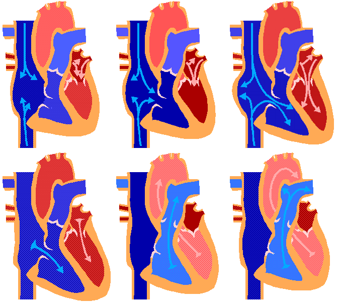

Heart is a pumping organ that keeps the blood continuously moving in the blood vessels. It is a blunt conical organ of about 12cm long and 9 cm broad. Its narrow apex is pointed downward and to the left. Heart is enclosed in a doubled walled sac, called pericardium. In between the two layers fluid filled space; the fluid is called pericardia l fluid and it prevents any friction between the heart walls and the surrounding tissues.
Human heart consists of four chambers. The two upper chambers are called atria (or auricles) and the two lower ones the ventricleTwo auricles are separated by an interatrial septum.
Two ventricles are separated by an interventricular septum Septum prevents the mixing of oxygenated blood and deoxygenated blood. The right auricle and right ventricle guarded by tricuspid valves. The left atrium and left ventricle are guarded by bicuspid valve. Both the valves are attached to chordae tendinae, the tough strands of connective tissue which in turn are attached to the papillary muscles of ventricles.
Heart is made of special muscles cells called cardiac muscles Bicuspid and tricuspid valves allows only unidirectional flow of blood i.e. from atria to ventricles Ventricles are far thicker walled than atria because ventricles pump out the blood with force to the different parts of the body, Left ventricles are far thicker walled than right ventricles as it is pumping the blood to different parts of the body a right ventricle is only up to lungs From right atrium the deoxygenated blood enters the right ventricle through tricuspid valves From right ventricle the deoxygenated blood is pumped out through pulmonary artery to lungs for oxygenation. Oxygenated blood from left atrium enters the left ventricles through bicuspid valve. From left ventricle, the oxygenated blood is pumped out into aorta, the largest artery which takes the blood to the body. In the tissues a capillary network allows the exchange of gases and food substances. Finally the deoxygenated blood flows through venules and then veins. This blood is returned to the right atrium through inferior and superior vena cava and inferior vena cava pour deoxygenated blood to the right atrium. Pulmonary veins from lungs pour oxygenated blood into left atrium. Right atrium pours deoxygenated blood into right ventricles right ventricles. Left atrium pours oxygenated blood into left ventricle.
CARDIAC CYCLE
1.contraction of atria.
right atrium recieves blood from vena ceva and pours into right ventricle. left atrium recieves blood from pulmonary vein and pours into left ventrical.
2.Contraction of both ventricles (Atria start relaxing):
from right ventricle, deoxygenated blood flows to the lungs through pulmonary artery.From left ventricle pours blood into aorta sends blood to the various parts of the body.

3 comments:
get me the real structure of heart
Hussain,
To see real structure of heart please click structure of heart.
Post a Comment